What Is TPLO Surgery? A Primer for New Surgeons
Quick answer
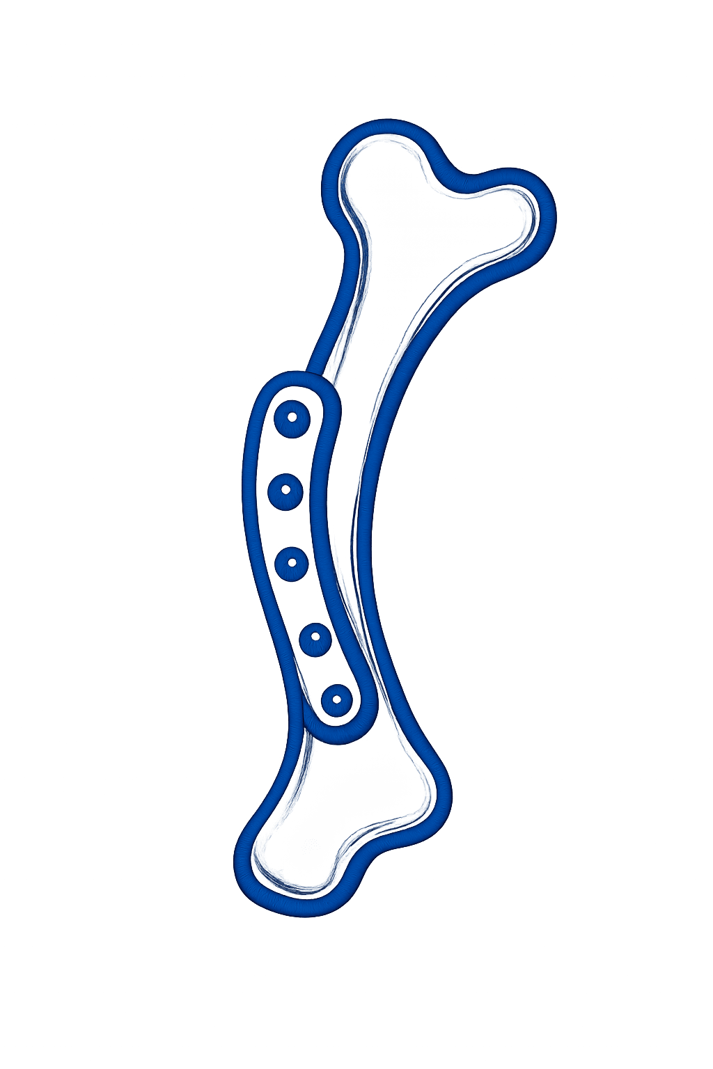
TPLO (tibial plateau leveling osteotomy) is the most widely used surgery for cranial cruciate ligament rupture in dogs. Instead of replacing the ligament, the surgeon cuts the top of the tibia in a curve, rotates it to level the sloped tibial plateau, and fixes it with a plate, so the knee stays stable when the dog bears weight.
Key takeaways
- TPLO treats cranial cruciate ligament (CrCL) disease by changing the geometry of the tibia, not by replacing the ligament.
- The key measurement is the tibial plateau angle (TPA); surgeons typically rotate the plateau to a post-op angle of about 5°.
- A curved (radial) osteotomy is made with a TPLO saw blade, the plateau segment is rotated a planned distance, and a locking plate holds it while the bone heals.
- Good outcomes start with a calibrated, well-positioned radiograph and a preoperative plan: TPA, blade size, osteotomy position, rotation, and plate.
Why dogs need TPLO: cranial cruciate ligament disease
The cranial cruciate ligament (CrCL, the canine equivalent of the human ACL) is one of the main stabilizers of the stifle (knee). In dogs it usually fails through progressive degeneration rather than a single athletic injury, which is why partial tears often become complete ruptures and why many dogs eventually rupture the ligament in the other knee too.
Without a working CrCL, the tibia slides forward relative to the femur every time the dog puts weight on the leg. That instability causes pain, lameness, joint inflammation, progressive osteoarthritis, and often a tear of the medial meniscus.
- Typical history: hind-limb lameness that may be sudden or intermittent, stiffness after rest, sitting with the affected leg kicked out.
- Exam findings: cranial drawer and a positive tibial thrust (tibial compression) test, stifle effusion, medial buttress (firm swelling on the medial joint), sometimes a meniscal click.
- Radiographs: joint effusion (loss of the infrapatellar fat pad), osteophytes in chronic cases. The ligament itself is not visible on radiographs.
The biomechanics: tibial thrust and the sloped plateau
The top of the canine tibia (the tibial plateau) slopes backward. When the dog bears weight, compressive force travels through the joint; because the plateau is sloped, part of that force becomes a forward-directed shear force on the tibia called cranial tibial thrust. A healthy CrCL resists it. A ruptured CrCL cannot.
Barclay Slocum's insight in developing TPLO was that you do not have to replace the ligament if you remove the slope. Rotate the plateau until it is nearly perpendicular to the tibia's functional axis, and weight bearing no longer pushes the tibia forward. In vitro work by Warzee and colleagues showed that leveling the plateau to about 6.5° neutralized cranial tibial thrust, which is why most surgeons aim for a post-op angle of roughly 5°.
Before surgery: the radiograph and the preoperative plan
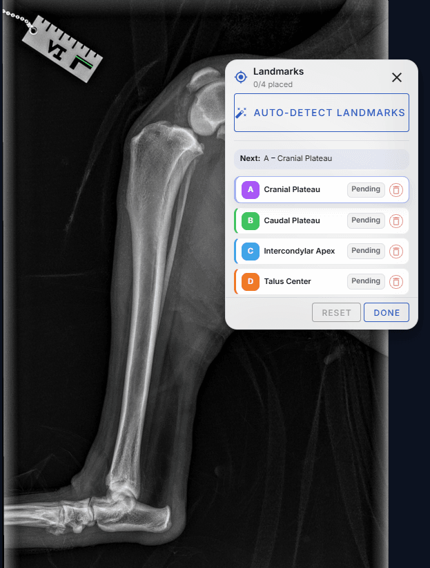
TPLO planning starts with a true mediolateral radiograph of the whole tibia, including the stifle and the tarsus (hock), with the femoral condyles superimposed and a calibration marker at the level of the bone. From that image the surgeon plans five things:
- Calibration: convert pixels to millimeters so every measurement is real. See radiograph positioning and calibration.
- TPA: place four landmarks (cranial and caudal plateau margins, the intercondylar eminence, and the center of the talus) and measure the angle.
- Saw blade and osteotomy position: choose a blade radius that fits the proximal tibia and position the curved cut, usually centered near the intercondylar eminence, while leaving enough tibial tuberosity.
- Rotation: calculate how far to rotate the plateau segment along the cut to reach the target angle. See TPLO rotation and saw blade selection.
- Plate: template a plate size that fits the proximal segment and the tibial shaft. See choosing and templating a TPLO plate.
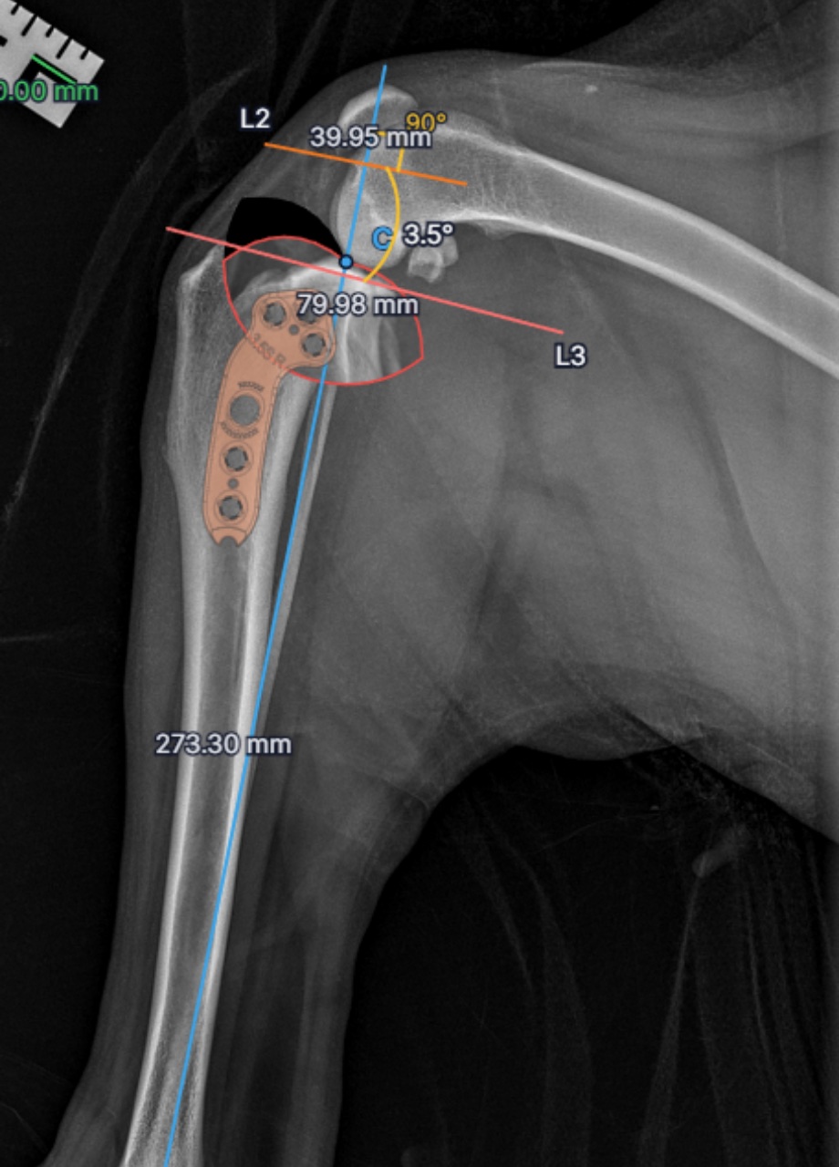
Done by hand with acetates and rotation charts, this takes around 30 minutes per case. For the full workflow, read how to plan a TPLO.
The surgery, step by step
Techniques vary between surgeons, but a typical TPLO follows this sequence:
- Joint assessment. The stifle is explored by arthrotomy or arthroscopy. Torn ligament remnants are debrided and the medial meniscus is inspected; a torn meniscus is treated, usually by partial meniscectomy.
- Approach and jig (optional). A medial approach exposes the proximal tibia. Some surgeons place a TPLO jig to hold alignment during the cut.
- Osteotomy. A curved, biradial TPLO saw blade makes the radial cut through the proximal tibia at the planned position.
- Rotation. The plateau segment is rotated along the cut by the planned distance, measured between reference marks on either side of the osteotomy, and held with a temporary pin.
- Fixation. A TPLO locking plate is applied to the medial tibia with screws in the proximal segment and the shaft.
- Post-op radiographs. Confirm the new TPA, the plate and screw positions, and that no screw enters the joint.
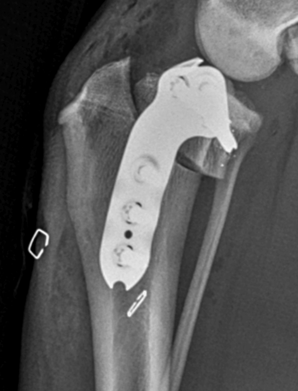
The post-op radiograph (shown at the top of this guide) is compared with the preoperative plan to confirm the plateau was leveled as intended.
Recovery and outcomes
Most dogs toe-touch within days and bear weight progressively over the following weeks. Activity is strictly restricted (leash walks only, no running, jumping or stairs) while the osteotomy heals, and recheck radiographs are usually taken around 8 weeks to confirm healing before activity is increased. Rehabilitation therapy is commonly used to rebuild muscle and range of motion.
TPLO is widely regarded as producing good to excellent long-term function in most dogs, including large and athletic breeds, which is a major reason it has become the most common CrCL procedure in referral practice.
Complications, and why planning matters
Reported complications include surgical site infection, tibial tuberosity fracture, fibular fracture, implant problems, patellar desmitis, and late meniscal tears. Several of these are influenced by decisions made during planning:
- Tibial tuberosity fracture is associated with osteotomy position and a thin remaining tuberosity. In a study of 468 TPLOs, planned cases had no tuberosity fractures in 172 surgeries versus 6.8% of free-hand cases (Collins and colleagues, 2014).
- Inadequate or excessive rotation leaves residual instability or over-corrects the plateau. A measured rotation distance, rather than an estimate, reduces that error.
- Poor implant fit (screws near the joint or the osteotomy) is easier to avoid when a true-size plate template has been checked before surgery.
Alternatives to TPLO
| Procedure | How it works | Notes |
|---|---|---|
| TPLO | Levels the tibial plateau with a curved osteotomy and plate | Most widely used; suits a wide range of dog sizes |
| TTA (tibial tuberosity advancement) | Advances the tibial tuberosity so the patellar ligament is perpendicular to the plateau | Different geometry; its own planning measurements |
| CBLO (CORA-based leveling osteotomy) | Levels the plateau with an osteotomy centered on the center of rotation of angulation | Used in some skeletally immature dogs and by preference |
| Lateral suture (extracapsular repair) | A synthetic suture outside the joint mimics the ligament | Traditionally used in smaller dogs; no osteotomy |
Frequently asked questions
What does TPLO stand for?
TPLO stands for tibial plateau leveling osteotomy: a curved cut (osteotomy) in the top of the tibia that lets the surgeon rotate and level the tibial plateau.
Does TPLO replace the cruciate ligament?
No. TPLO changes the angle of the tibial plateau so that weight bearing no longer drives the tibia forward. The stifle becomes functionally stable without a replacement ligament.
What angle is the plateau rotated to in a TPLO?
Most surgeons target a post-operative tibial plateau angle of about 5°, based on in vitro evidence that leveling to around 6.5° neutralizes cranial tibial thrust.
How long does TPLO recovery take?
Bone healing usually takes around 8 to 12 weeks, with strict activity restriction until recheck radiographs confirm healing, followed by a gradual return to normal activity.
What do you need to plan a TPLO?
A calibrated, true lateral radiograph of the whole tibia, the tibial plateau angle, a chosen saw blade radius and osteotomy position, the rotation distance, and a plate template. osAlign does all of these on one screen.
References
- American College of Veterinary Surgeons (ACVS). Cranial Cruciate Ligament Disease. https://www.acvs.org/small-animal/cranial-cruciate-ligament-disease
- Kim SE, Pozzi A, Kowaleski MP, Lewis DD. Tibial osteotomies for cranial cruciate ligament insufficiency in dogs. Vet Surg. 2008;37(2):111-125. https://pubmed.ncbi.nlm.nih.gov/18251804/
- Collins JE, Degner DA, Hauptman JG, DeCamp CE. Benefits of pre- and intraoperative planning for tibial plateau leveling osteotomy. Vet Surg. 2014;43(2):142-149. https://pubmed.ncbi.nlm.nih.gov/24491234/
- Johnston SA, Tobias KM, eds. Veterinary Surgery: Small Animal. 2nd ed. St. Louis: Elsevier; 2018.
- Slocum B, Slocum TD. Tibial plateau leveling osteotomy for repair of cranial cruciate ligament rupture in the canine. Vet Clin North Am Small Anim Pract. 1993;23(4):777-795. https://pubmed.ncbi.nlm.nih.gov/8337790/
- Warzee CC, Dejardin LM, Arnoczky SP, Perry RL. Effect of tibial plateau leveling on cranial and caudal tibial thrusts in canine cranial cruciate-deficient stifles: an in vitro experimental study. Vet Surg. 2001;30(3):278-286. https://pubmed.ncbi.nlm.nih.gov/11340560/
- Kowaleski MP, Apelt D, Mattoon JS, Litsky AS. The effect of tibial plateau leveling osteotomy position on cranial tibial subluxation: an in vitro study. Vet Surg. 2005;34(4):332-336. https://pubmed.ncbi.nlm.nih.gov/16212587/
- Bergh MS, Peirone B. Complications of tibial plateau levelling osteotomy in dogs. Vet Comp Orthop Traumatol. 2012;25(5):349-358. https://pubmed.ncbi.nlm.nih.gov/22534675/
For educational and planning support only. Surgeon judgment governs all clinical decisions.
Related guides
Plan your next TPLO in under 60 seconds
AI places the landmarks, the TPA is calculated instantly, and every measurement is reported to 0.1 mm. 30-day free trial with up to 5 cases, no credit card required.
Start free trial